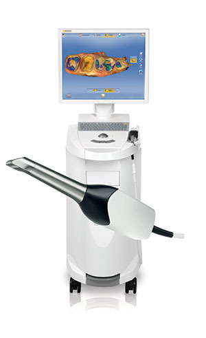

Analyses involving a scanning electron microscope can be inaccurate if the angle of the specimen is not correct. Stereomicroscope techniques require a transverse section of the crown and tooth to measure misfit, but this can cause deformations. A marginal fit of between 25 and 40 μm for cemented restorations has been suggested as a clinical goal, but these levels are rarely achieved.ĭifferent methods have been used to evaluate marginal fit, including stereomicroscopy, scanning electron microscopy, optical microscopy, and micro-computed tomography (μ-CT). Still others argue that a clinically acceptable fit should be ≤75 μm. Some studies indicate that a marginal fit ≤120 μm is clinically acceptable, but others have concluded that a marginal fit ≤100 μm is more suitable. No consensus yet exists regarding clinically acceptable marginal fit. Marginal fit is an important predictor of the clinical success of ceramic restorations. This technique was developed to reduce processing inconsistencies associated with conventional sintering, to avoid microcracking, and to improve mechanical stability. For this technique, ingots of lithium disilicate are subjected to heat-pressing that uses a pneumatic ram within a porcelain furnace to press ceramic material into the mold. This technique can cope with errors in preparation and design better than CAD/CAM systems. After milling, the precrystallized block crowns must be subjected to a heat crystallization process to achieve definitive strength.ĭental laboratories can also use the lost-wax technique to generate ceramic crowns. However, methods for obtaining the cast and milling the crown are different for the Cerec and E4D systems. These systems allow clinicians to fabricate restorations during a single patient visit by using intraoral optical impressions and in-office milling. Blocks of lithium disilicate are available for in-office chairside CAD/CAM systems, including Cerec (Sirona Dental Systems GmbH), E4D (Planmeca-E4D), and others. Lithium disilicate crowns can be fabricated in the dental office or the dental laboratory. Computer-aided design and computer-aided manufacturing (CAD/CAM) procedures are available in many dental practices, dental laboratories, and production centers. Crowns can be produced from ceramic materials by using a number of different techniques, each of which can reduce both chair time and laboratory processing time compared with conventional fixed prostheses. Ceramic restorations exhibit enamel-like color and translucency. Increasing patient demand for esthetic dental restorations and the improved physical properties of ceramic materials have made ceramic systems popular for dental restorations. The Cerec 3D Bluecam scanner chairside CAD/CAM system and the heat-pressing technique provided more accurately fitting crowns than the E4D Laser scanner chairside CAD/CAM system. Lithium disilicate crowns fabricated by using the Cerec 3D Bluecam scanner CAD/CAM system or the heat-pressing technique exhibited a significantly smaller vertical misfit than crowns fabricated by using an E4D Laser scanner CAD/CAM system. Both types of horizontal misfit (underextended and overextended) were 49.2% for heat-pressing, 50.8% for Cerec, and 58.8% for E4D.


The percentage of crowns with a vertical misfit <75 μm was 83.8% for Cerec and heat-pressing, whereas this value was 65% for E4D. The mean values of vertical misfit were 36.8 ☑3.9 μm for the heat-pressing group and 39.2 ☘.7 μm for the Cerec group, which were significantly smaller values than for the E4D group at 66.9 ☓1.9 μm ( P =.046). Data were statistically analyzed by 1-way ANOVA, followed by the Tukey honestly significant difference test (α=.05). Each crown was fixed to the cast and scanned with micro-computed tomography to obtain 52 images for measuring the vertical and horizontal fit. Three fabrication techniques were used: digital impressions with Cerec 3D Bluecam scanner with titanium dioxide powder, followed by milling from IPS e.max CAD for Cerec digital impressions with E4D Laser scanner without powder, followed by milling from IPS e.max CAD for E4D and fabrication from IPS e.max Press by using the lost-wax and heat-pressing techniques. Lithium disilicate crowns were fabricated to fit an in vitro cast of a single human premolar. The purpose of the study was to evaluate with micro-computed tomography the marginal fit of lithium disilicate crowns fabricated with different chairside CAD/CAM systems (Cerec or E4D) or the heat-pressing technique. No consensus exists concerning the acceptable ranges of marginal fit for lithium disilicate crowns fabricated with either heat-pressing techniques or computer-aided design and computer-aided manufacturing (CAD/CAM) systems.


 0 kommentar(er)
